
The Tönnis angle is determined by drawing three lines:Ī horizontal line connecting the base of the acetabular teardropsĪ horizontal line parallel to line 1, running through the most inferior point of the sclerotic acetabular SourcilĪ line extending from most inferior to the lateral margin of the acetabular Sourcil. Appropriate film exposure and contrast should enable delineation of the acetabular walls and the fat pads around the hip joint (gluteal, obturator and iliopsoas). Radiographic contrast is defined as the density difference between neighbouring areas on a plain radiograph.

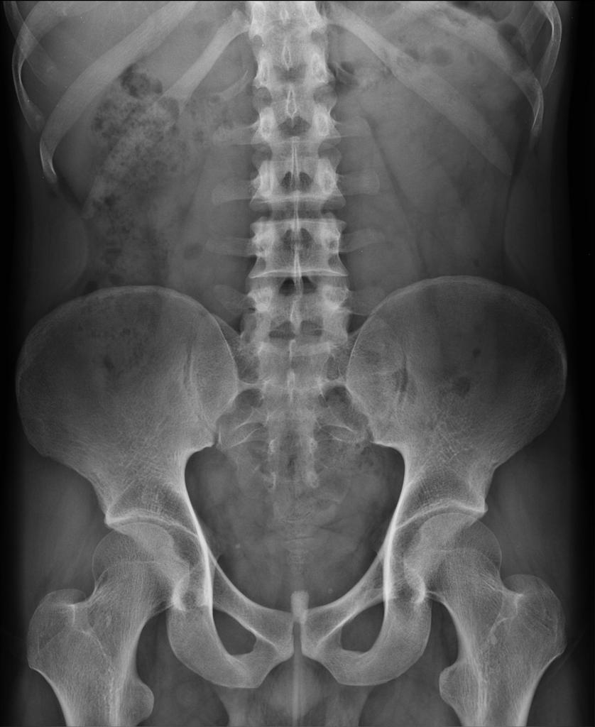
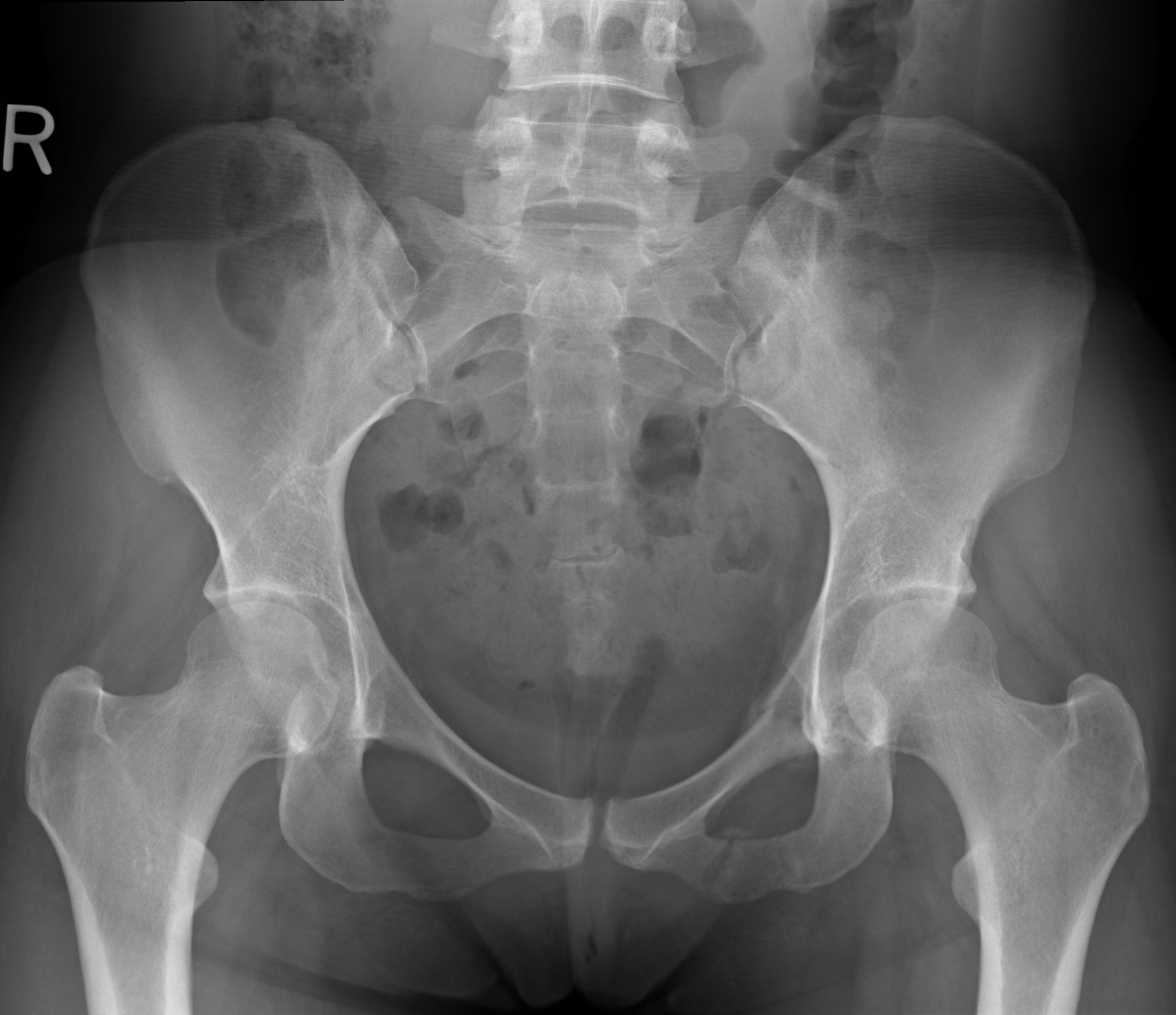
The exposure of the radiograph is related the concentration of photons absorbed by the film, or digital detector, at the time the radiograph was taken. Acquisition of the radiographic image with the patellae facing anteriorly can lead to over-estimation of the true neck-shaft angle. Rotation of the lower limbĪpproximately 15° of internal rotation of the hips, such that the greater and lesser trochanters are parallel to the floor, maximises the length of the femoral neck and enables a more accurate neck-shaft angle measurement. The distance between the sacro-coccygeal junction and the superior end of the symphysis should be between 2 and 3 cm in males and between 2 and 6 cm in females (Fig. 1).Ī plain AP radiograph should have neutral tilt, which may require correction of lumbar lordosis. The central sacral line and the tip of the coccyx should also align with the symphysis pubis (Fig. In the absence of pathology, the two obturator foramen, ischial spines, greater and lesser trochanters and femoral heads should be symmetrical.

Pelvis rotationĪn acceptable plain radiograph of the pelvis should be obtained with pelvis in a neutral position. In addition, the following factors should be considered. Images should allow for visualisation of the entire pelvis including the iliac crests, sacrum, sacroiliac joints, pubic and ischial rami, as well as the necks of the femora and the lesser and greater trochanters. When initially reviewing a plain radiograph of the pelvis, the radiograph should be assessed for adequacy. It can also be obtained in a weight-bearing manner, which may accentuate any arthritic changes, and give an indication of limb length discrepancy. The AP pelvis X-ray can be performed in a supine position, with efforts made to control the pelvic tilt and rotation, enabling similar radiographs to be obtained in every patient. Acetabular and femoral parameters that are assessed on plain radiographs are then described.Ī plain antero-posterior (AP) radiograph of the pelvis facilitates an assessment of each hip joint on an individual basis, as well as allowing for a comparison to be made to the contra-lateral hip joint. This article aims to summarise the most important aspects of the assessment of plain radiographs performed on the young adult hip joint. This paper begins by describing the parameters that potentially impact the quality of antero-posterior (AP) and lateral radiographs of the hip, and the variations in lateral radiographs that can be used. Radiographic examination remains the mainstay of the initial assessment however, common parameters are required to assist in the formation of accurate diagnoses and appropriate management plans including appropriate further imaging. An enhanced awareness of the presence of structural disorders of the hip, such as developmental dysplasia of the hip and femoroacetabular impingement (FAI), has fuelled an evolution in the assessment of patients with hip pain and enhanced our ability to diagnose patients, even in cases where there are mild structural abnormalities.


 0 kommentar(er)
